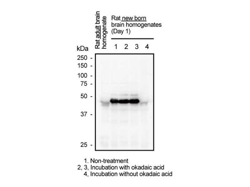
Phosphorylation of signal transduction molecules Smad3, can be an important information for understanding of various biological functions of transforming growth factor (TGF)-β. TGF-β type I receptor and mitogen activated protein kinase (MAPK) phosphorylate Smad3 at the C-terminals and at linker (middle) regions respectively. Mitogenic signaling induced by HGF, EGF and inflammatory cytokines is mediated by Smad3 phosphoisoform phophorylated serine located in linker region. This monoclonal antibody against for Smad3L (S213) recognizes the part of phosphorylated serine (Ser213) located in linker region of Smad3 specifically. It can be used for western blotting, immunoprecipitation and immunohistochemistry. It is applicable for enzyme biochemical analysis of Smad3 signaling and makes it possible to visualize the intermolecular reaction of phosphorylated Smad3 signaling in human tissues and monitor them in real-time. Thus, analysis of Smad3 signaling with this antibody is expected to be widely applied to cancer research and fibrosis research, and to contribute to understanding the wide variety of life phenomena mediated by phosphoisoforms of Smad3. For research use only, not for use in diagnostic procedures.
- application:
- IHC, WB, IP
- Catalog number:
- 10387-S
- clone:
- 5A11
- concentration:
- Please see datasheet
- Datasheet:
- formulation:
- Lyophilized product in PBS containing 1% BSA and 0.05% NaN3
- immunogen:
- Synthetic peptide of phosphorylated Smad3L (S213)
- isotype:
- IgG1
- MSDS:
- notes:
- For research use only, not for use in diagnostic procedures.
The datasheet for this product (see above) is intended to serve as an example only. Please refer to the datasheet provided with the antibody for precise details. - Other names:
- Please see datasheet
- Protocol:
- size:
- 5 µg
- storage:
- Lyophilized product, 5 years at 2 - 8 °C; Solution, 2 years at -20 °C
- Host:
- Mouse


